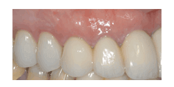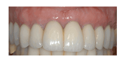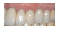
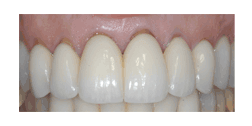
Even after recent crown insertion, patients often present with exposed crown margins. I am frequently surprised when I see ample width and thickness of keratinized tissue, associated with gingival recession. This dilemma can be predictably resolved with root coverage techniques. Using a subepithelial connective tissue graft and microsurgical techniques can result in coverage of the crown margin with little to no discomfort or swelling. Below are pre-operative photos showing the gingival response one year after crown insertion. The post-operative photos were taken at four months after surgery.
Below are pre-op photos showing the gingival response one year after crown insertion
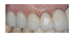
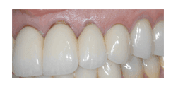
These post-op photos were taken at 4 months after surgery