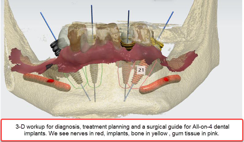
Recent advances in imaging now allow for more predictable treatment outcomes. The 3D CBCT (three-dimensional cone beam computed tomography) scanner allows for accurate visualization of anatomical structures such as the structure of the jaw bone, and nerve location which is critical for proper diagnosis and rendering treatment in a safe manner.
Traditional dental X-rays are two-dimensional and do not provide the amount of information necessary for many dental implant and periodontal surgical procedures.
To offer our patients the advantages of the best technology available, Dr. Kissel has had installed the JMorita Veraviewepocs 3D R100. The primary reasons for selecting this scanner are its ability to limit exposure to radiation and its excellent resolution.
Many CBCT scanners use a cylindrical field of view which takes images that include areas outside the region of interest. This increases the dosage of exposure. By abandoning the typical cylindrical field of view with a new convex triangular shape (which closely matches the dental arch form), the dosage of radiation is significantly reduced.
The high resolution of this scanner is a dramatic improvement over many existing scanners. Obtaining a very clear view of the structures of interest will result in precise, detailed, safe, and predictable treatment.
To learn how CBCT technology can be used in treatment planning your dental needs please don’t hesitate to ask Dr. Kissel.
